Hip
Coverage from top of iliac crest to below lesser trochanter.
3 plane recons and 3D
Knee
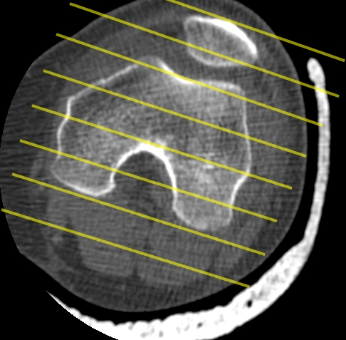
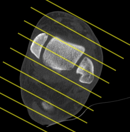
Coronals parallel to posterior condylar line - sagittals perpendicular to that
ARTHROGRAM PROCEDURE
20-30cc of undiluted Niopam 200-300. Exercise to distribute
Compression bandage over patella. Knee flexed 20 degrees
Parameters kV=140 mA=135 <1mm slice thickness. Low pitch
Axial scan through contrast region.
Use axial data set to obtain coronal and sagittal reformats. Align with posterior margin of the femoral condyles as shown

Ankle & Foot CT
Start with ablock of axial images from 3cm above the tibial plafond to include all phalanges.
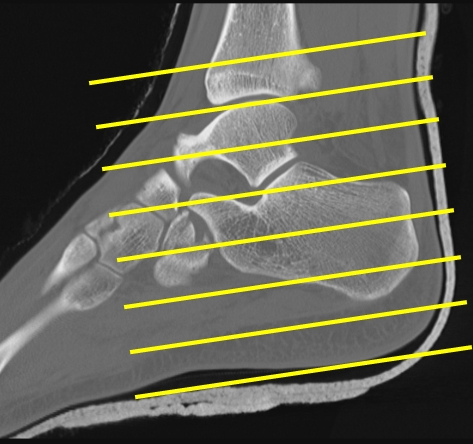
Hindfoot Recons
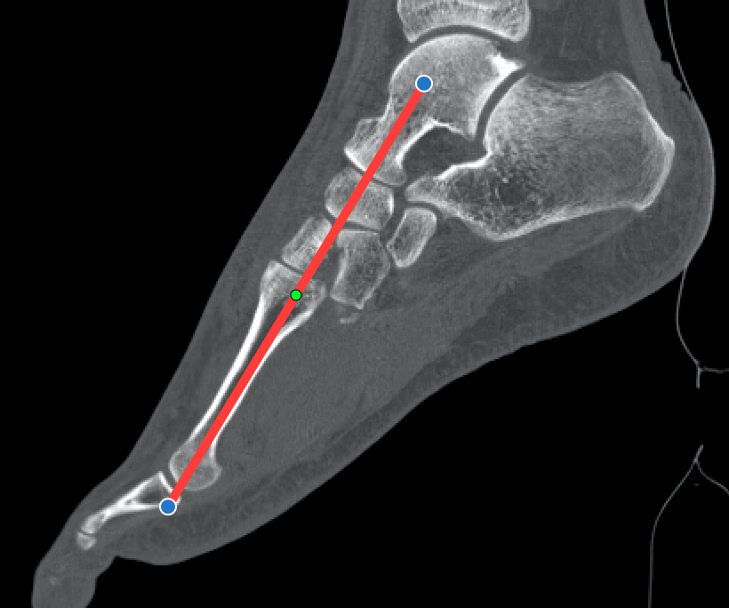
Forefoot Recons
Forefoot reconstructions require careful planning. Start by loading a simple MPR into the worstation. Sagittal recons are aligned perpendicular to the short axis of the forefoot as in figure 1 to the right. Axial recons (long axis) need to be double obliques, first tilted as in image 2 to take account of foot position, then aligned along the metatarsal bones as in image 3. If the request refers to a specific metatarsal or digit (eg Great Toe) then align along that metatarsal. The final set of true coronal (short axis) recons are then perpendicular to the sagittals


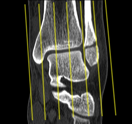
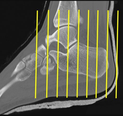
Orientate coronal recons along the anterior malleolar line as in the image immediatly to the right. The anterior margin of the talus can also be used as the two are similar. Sagittal recons are perpendicular to this but also tilted to align along the long axis of the tibia. See the 2nd image adjacent. Axial recons are aligned along the long axis of the talus as shown in image 4.
Midfoot Recons eg Lisfranc fracture
Reconstructions are as per conventional forefoot recons outlined above but use the 2nd metatarsal for sagittal alignment